


Description
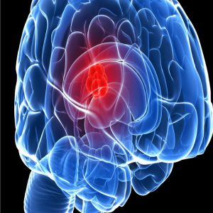
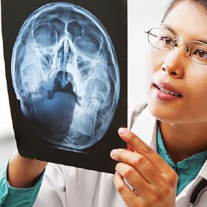
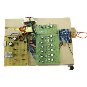
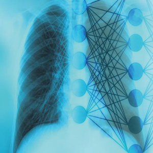
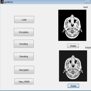
Lung Cancer detection Using Matlab
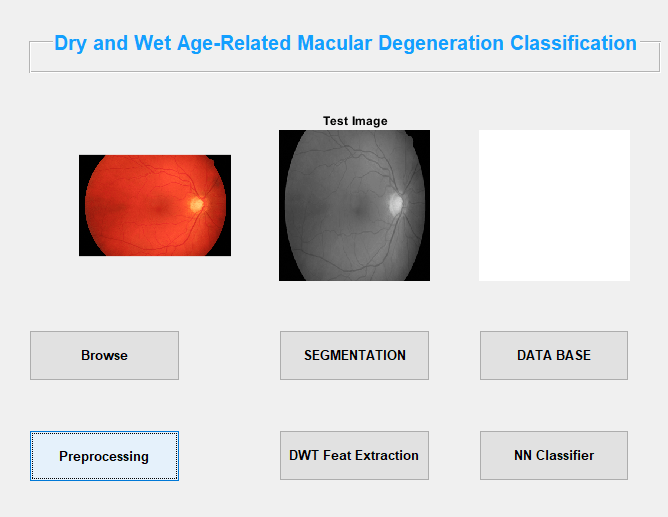
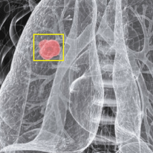
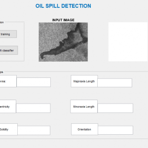
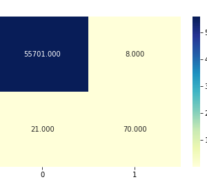
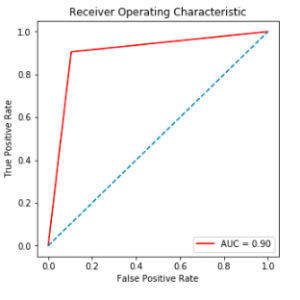
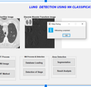
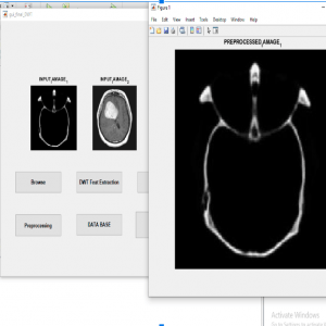
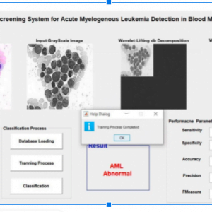
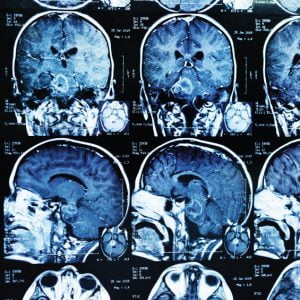
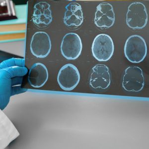
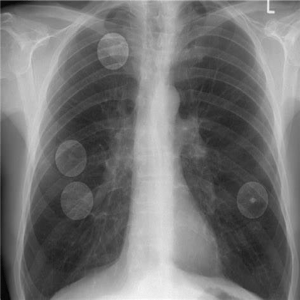
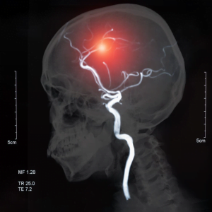
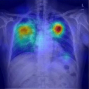
Lung diseases are the disorders that affect the lungs and the organs that allow us to breathe and it is the most common medical condition worldwide, especially in India. The diseases such as pleural effusion and normal lung are detected and classified in this work. This paper presents a computer-aided classification Method in Computer Tomography (CT) Images of lungs developed using NN. The purpose of the work is to detect and classify lung diseases by effective feature extraction through Dual-Tree Complex Wavelet Transform and GLCM Features. The entire lung is segmented from the CT Images and the parameters are calculated from the segmented image. The parameters are calculated using GLCM. We Propose and evaluate the Network designed for the classification of ILD patterns. The parameters give the maximum classification accuracy. After the result, we propose clustering to segment the lesion part from the abnormal lung.
OVERVIEW AND SCOPE:
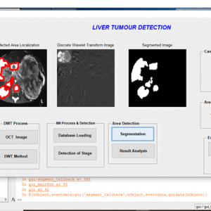
The main objective of this project is to identify cancer in an? lung image using deep learning? technique along with image processing .if the person is infected with a lung tumor.it will easily identify with help of a ct-scan image .and? then classified into two stages normal and abnormal .it shows where it is affected in the particular area of the lung. In this we used DT-CWT, GLCM FEATURE EXTRACTION to detect the lung tumor.
EXISTING SYSTEM
- K means clustering?
- Wavelet and Principal component analysis
- KNN classifier
Drawbacks:
- Difficult to get accurate results
- Not applicable for multiple images for lesion segmented in a short time
- Poor discriminatory power? and less classification? accuracy
Proposed Method:
- Preprocessing?
- Dwt
- Glcm feature extraction
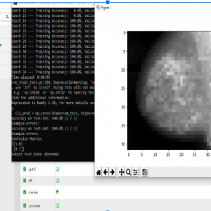
- Neural network
Advantages:
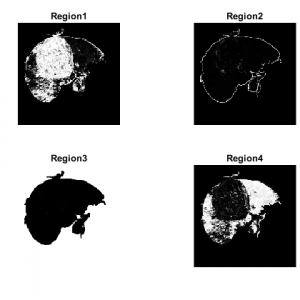
- The segmentation algorithm Proves to be simple and effective
- Grayscale Co-occurrence matrix performed well in BPN
- Better texture and edge representation?
- Segmentation provides better clustering efficiency
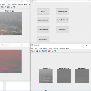
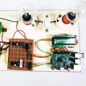
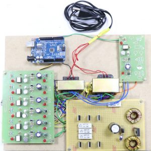
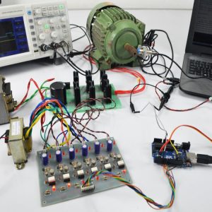
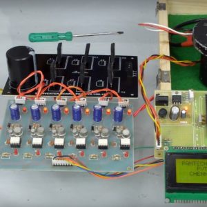
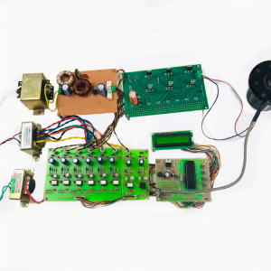
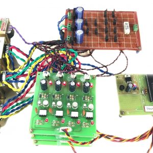
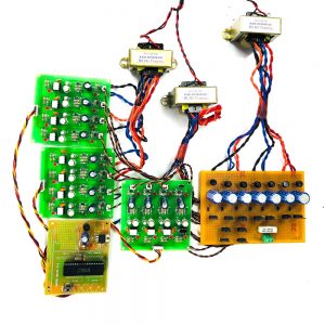
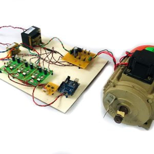
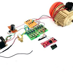
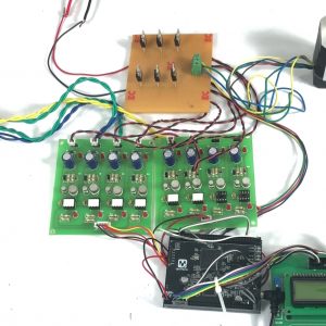
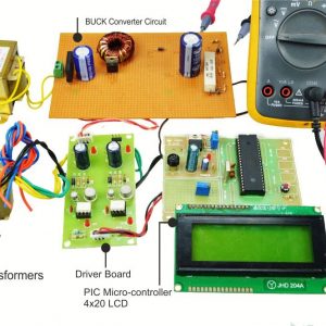
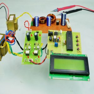
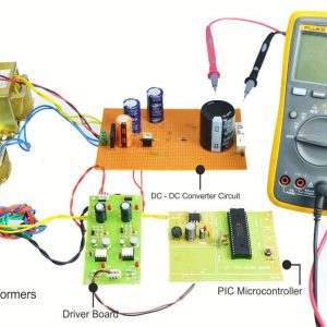
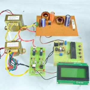
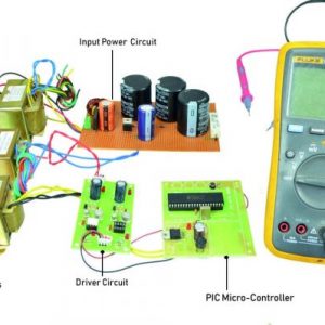
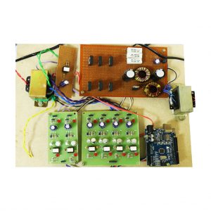
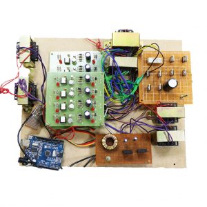
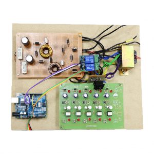
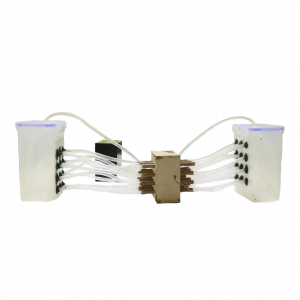
BLOCK DIAGRAM

PREPROCESSING
Digital image processing deals with?manipulation of digital images through a digital computer. It is a subfield of signals and systems but focus particularly on images. DIP focuses on developing a computer system that is able to perform?processing?on an?image. The input of that system is a digital?image?and the system process that?image?using efficient algorithm
It allows a much wider range of algorithms to be applied to the input data and can avoid problems such as the build-up of noise and distortion during processing.
- Importing the image via image acquisition tools;
- Analysing and manipulating the image;
- Output in which result can be altered image
Image Pre-processing?is a common name for operations with?images?at the lowest level of abstraction. Its input and output are intensity?images.? The aim of?pre-processing?is an improvement on the? image? or data that suppresses unwanted distortions or enhances some images? features important for further processing.
Lung Cancer detection Using Matlab
DIGITAL IMAGE PROCESSING
CLASSIFICATION OF IMAGES
There are 3 kinds of pix applied in Digital Image Processing. They are
- Binary Image
- Gray Scale Image
- Colour Image
Binary picture
A binary photograph is a virtual photo that has most effective? viable values for every pixel.? Typically the two colorings used for a binary image are black and white despite the truth that any? colors may be used.? The color used for the item(s) in the photograph is the foreground color while the rest of the picture is the historic past coloration.
Binary pix are also called bi-stage or -stage. This approach is that every pixel is stored as a single bit (zero or 1). This name black and white, monochrome or monochromatic are regularly used for this idea, however may also additionally designate any pics that have best one pattern in keeping with pixel, together with grayscale photos
?Binary pics often upward push up in virtual image processing as masks or because the result of positive operations collectively with segmentation, thresholding, and dithering. Some input/output gadgets, which include laser printers, fax machines, and bi-level laptop shows, can most effectively manage bi-degree pix.
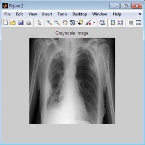
Grayscale images
A grayscale Image is a virtual photo that is a picture in which the price of every pixel is a single sample, this is, it consists of satisfactory depth information. Images of this type, also referred to as black-and-white, are composed solely of sun shades of gray(0-255), various from black(0) on the weakest depth to white(255) at the maximum effect.
Grayscale snapshots are extremely good from one-bit black-and-white pix, which in the context of laptop imaging are pix with only the 2 sun sunglasses, black, and white (additionally known as bi-diploma or binary photos). Grayscale snapshots have many sun shades of grey in them. Grayscale photos are also referred to as? ? monochromatic, denoting the absence of any chromatic version.
Grayscale pix are frequently the quit end result of measuring the intensity of moderate at every pixel in a single band of the electromagnetic spectrum (e.G. Infrared, seen mild, ultraviolet, and lots of others.), and in such instances they will be monochromatic proper at the same time as simplest a given frequency is captured. But moreover, they may be synthesized from a whole color picture; see the phase approximately changing to grayscale.
Colour picture
A (digital) colour image is a virtual photograph that includes shade records for every pixel. Each pixel has a specific rate that determines its appearing shade. This fee is certified by means of manner of 3 numbers giving the decomposition of the color inside the 3 number one sun shades Red, Green, and Blue. Any shade seen by the human eye may be represented in this manner. The decomposition of color inside the 3 number one hues is quantified through some of them amongst 0 and 255. For instance, white may be coded as R = 255, G = 255, B = 255; black can be referred to as (R,G,B) = (0,0,0); and say, amazing pink may be : (255,0,255).?
In great phrases, a photo is an intensive -dimensional array of color values, pixels, each of them coded on three bytes, representing the 3 primary shades. This allows the image to encompass a complete of 256x256x256 = 16. Eight million particular shades. This approach is likewise referred to as RGB encoding, and is particularly tailor-made to human imaginative and prescient
From the above determine, sun shades are coded on 3 bytes representing their decomposition on the 3 primary colorings. It sounds obvious to a mathematician to right away interpret colors as vectors in a three size area in which every axis stands for one of the number one hues. Therefore we are able to gain a maximum of the geometric mathematical requirements to cope with our colors, which consist of norms, scalar product, projection, rotation, or distance.Fig.1 Hue Saturation
Process of RGB SCALE Image
The identification of devices in an picture and this method may additionally possibly begin with photograph processing techniques collectively with noise removal, observed via the usage of (low-diploma) feature extraction to find outlines, areas and probable regions with sure textures.
The smart bit is to interpret collections of those shapes as single devices, e.G. Vehicles on an avenue, boxes on a conveyor belt, or cancerous cells on a microscope slide. One motive for this AI problem is that an item can seem very one-of-a-type while viewed from unique angles or underneath amazing lighting. Another problem is finding out what skills belong to what item and which can be information or shadows and so on. The human visible device plays these responsibilities specifically unconsciously however a laptop calls for skillful programming and masses of processing strength to method human commonplace overall performance. Manipulation of records inside the shape of an photograph through several viable strategies. An image is generally interpreted as a -dimensional array of brightness values, and is maximum familiarly represented via way of such patterns as the ones of a photographic print, slide, tv display, or movie show display. An image may be processed optically or digitally with a laptop.
IMAGE ACQUISITION
Image Acquisition is collecting a digital photograph. To collect this requires a picture sensor and the functionality to digitize the sign produced thru the sensor. The sensor might be monochrome or coloration TV camera that produces an entire photo of the trouble area each 1/30 sec. The photograph sensor may also be a line test virtual digicam that produces a single photo line at a time. In this situation, the gadgets move beyond the road.?
IMAGE ENHANCEMENT
Image enhancement is most of the best and maximum appealing regions of digital image processing. Basically, the concept in the back of enhancement strategies is to perform detail that is obscured, or clearly to spotlight certain features of exciting an picture. A familiar instance of enhancement is at the same time as we grow the assessment of a photograph due to the fact? it appears higher.? It is essential to remember the fact that enhancement is a completely subjective place of photo processing.
IMAGE RESTORATION
Image recuperation is a place that also deals with enhancing the appearance of an image. However, not like enhancement, which is subjective, photograph recuperation is the goal, in the experience that restoration techniques will be predisposed to be based on mathematical or probabilistic fashions of picture degradation.
Conclusion:
Lung cancer is the most dangerous and widespread in the world according to stage the discovery of the cancer cells in the lungs, this gives us the indication that the process of detection this disease plays a very important and essential role to avoid serious stages and to reduce its percentage distribution in the world. To obtain more accurate results we divided our work into three stages: Image Enhancement stage, Image Segmentation stage, and Features Extraction stage. Lung Nodule Detection in CT Scans is an active area of research that is continuously emerging and there are many enhancements that can be included to make it more efficient.
Lung Cancer detection Using Matlab
REFERENCES
[1] American Cancer Society, ?Cancer Statistics, 2005?, CA: A Cancer Journal for Clinicians, 55: 10-30, 2005, ?http://caonline.amcancersoc.org/cgi/content/full/55/1/10?.?
[2] D. Lin and C. Yan, ?Lung nodules identification rules extraction with the neural fuzzy network?, IEEE, Neural Information Processing, vol. 4, (2002).?
[3] A. El-Baz, A. A. Farag, PH.D., R. Falk, M.D. and R. L. Rocco, M.D., ?detection, visualization, and identification of lung abnormalities in chest spiral CT scans: phase I?, Information Conference on Biomedical Engineering, Egypt (2002).
?[4] B.V. Ginneken, B. M. Romeny and M. A. Viergever, ?Computer-aided diagnosis in chest radiography: a survey?, IEEE, transactions on medical imaging, vol. 20, no. 12 (2001).?
[5] Beucher, S. and Meyer, F., ?The Morphological Approach of Segmentation: The Watershed Transformation,? Mathematical Morphology in Image Processing, E. Dougherty, ed., pp. 43-481, New York: Marcel Dekker, 1992.?
[6] Nguyen, H. T., et al ?Watersnakes: Energy-Driven Watershed Segmentation?, IEEE Transactions on Pattern Analysis and Machine Intelligence, Volume 25, Number 3, pp.330-342, March 2003.
?[7] Suzuki K., et al.,?False-positive Reduction in Computer-aided Diagnostic Scheme for Detecting Nodules in Chest Radiographs by Means of Massive Training Artificial Neural Network?, Academic Radiology, 12, No 2, February 2005, pp. 191-2016
?


Customer Reviews
There are no reviews yet.