

Description
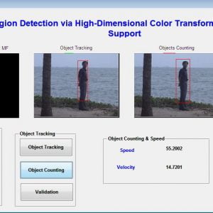
Oral Cancer Detection using Image Processing
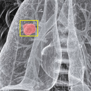
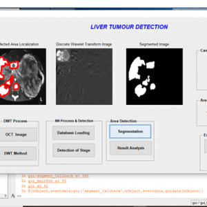
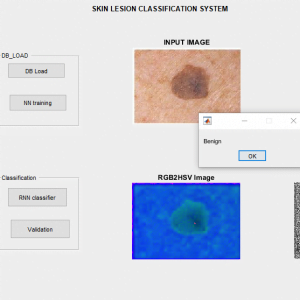
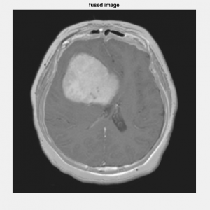
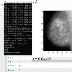
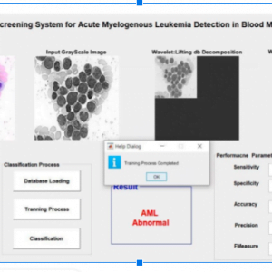
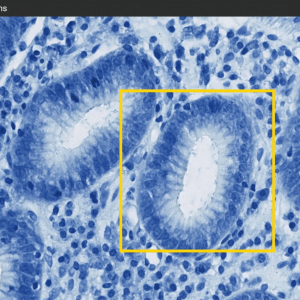
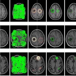
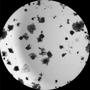
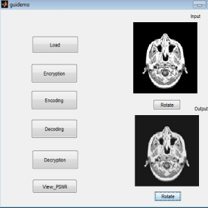
The tumor occurs in salivary glands, tonsils, and also in the neck, head, face, and oral cavity. There are various diagnosis methods to find oral cancer such as the biopsy method in which a small sample of tissues is removed from a part of the body and tested in the microscope. And some? screening methods. But the drawback is we cannot actually clearly detect the tumor of cancer cells as well as we couldn’t classify how many cells are affected by cancer so in this paper we are going to detect and classify the affected cancerous cell in the oral region by digital Image processing techniques feature extraction enables clear visualization of cancer-affected areas. Here we use the firefly algorithm to detect the cancer tumor in the MRI image. And Backpropagation neural network to classify the cancer cells accurately the project is carried out using a mat lab program. Oral Cancer Detection using Image Processing
INTRODUCTION
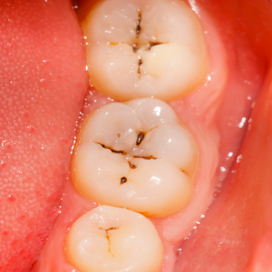
The tumor occurs in salivary glands, tonsils, and also in the neck, head, face and oral cavity people die around 1,30000 in a year? with this cancer? According to the American cancer society men face twice the risk of developing oral cancer as women, men who are over age 40 face a greater risk, over 25% of all oral cancers occur in people who do not smoke who drink alcohol occasionally 75% of oral cancer are due to use of tobacco and excessive alcohol consumption on other factors such as poor oral hygiene poor nutrition and by chronic infection caused by bacteria and virus.? Oral cancer occurs in the mouth and throat Oral cancer is fairly common and Very curable if found and treated at an early stage. Oral cancer is also known as mouth cancer. This arises in any of the tissues in the mouth. There are several types of oral cancers, but around 90% are squamous cell carcinoma originating in the tissues that line the mouth and lips. Oral or mouth cancer most commonly involves the floor of the mouth cheeks, gums, lips, or palate roof of the mouth. Most oral cancers look very similar under the microscope and are called squamous cell carcinoma The Signs and symptoms are such as tongue and lips usually painless at initial, the Burning sensation will occur if the tumor gets advanced, Additional Problems are such as swallowing difficulty, mouth sores, etc.
EXISTING SYSTEM
Oral cancer detection using tumor-based marker using a watershed algorithm. The technique used in this paper is Orthopantomogram. And the watershed algorithm is proposed to preserve the edge details as well as prominent ones to identify tumors in dental radiographs. Marker Controlled Watershed segmentation is used to segment tumors because watershed on images leads to over-segmentation even though it is preprocessed.
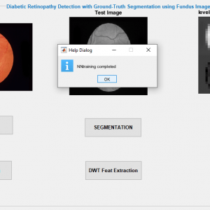
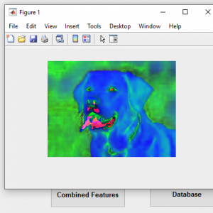
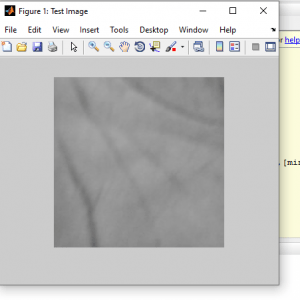
PROPOSED SYSTEM
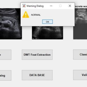
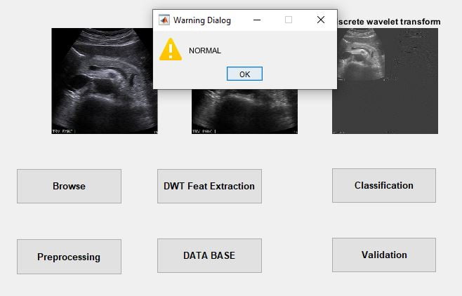
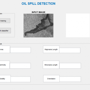
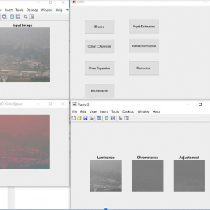
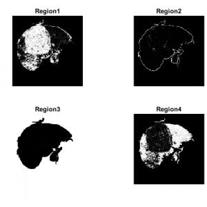
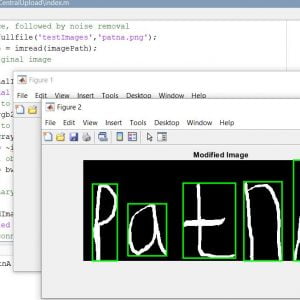
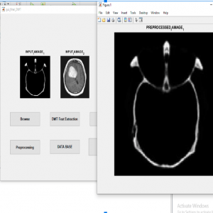
Proposed methodology includes enhancement of images we used salt and pepper noise, segmentation of cells are used for thresholding but that we can find the accurate results and find very easily tumor by partition, features extraction, and finally the classification with the Backpropagation neural networks.


Customer Reviews
There are no reviews yet.