

Description
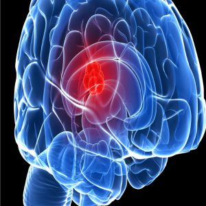
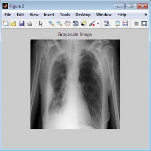
Lung Nodule Detection in X ray Images using Image Processing
Abstract
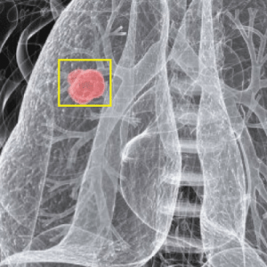
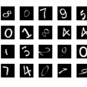
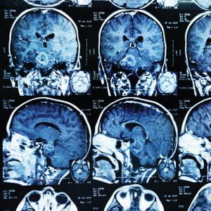
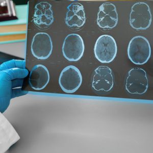
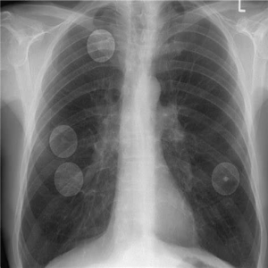
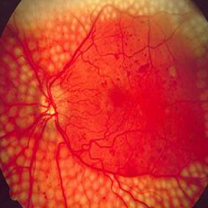
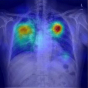
In this project, a novel procedure to apply deep learning techniques to medical image classification was proposed. With the increasing popularity of Chest X rays, fully manual diagnosis of lung nodules puts a burden on the radiologists who need to spend hours reading through Lund nodule images to identify regions of Interest (ROIs) to schedule follow-ups. Accurate computer-aided diagnosis of lung cancer can effectively reduce their workload and help train new radiologists. However, lung nodule detection is challenging because of the varying size, location, shape, and density of nodules. Many studies have approached this problem using image-processing techniques with the intention of developing an optimal set of features. Convolution neural network has demonstrated to learn discriminative visual features automatically and has beat many state-of-art algorithms in image-processing tasks, such as pattern recognition, object detection, segmentation, etc. In this report, we evaluate the feasibility of implementing deep learning algorithms for lung cancer diagnosis with the Lung Image Database Consortium (LIDC) database.. The performance of our best model is comparable with the state-of-art results in the lung nodule detection task. Lung Nodule Detection in X ray Images using Image Processing
System Analysis
Existing Systems
- Principal Component Analysis
- DCT and shape features
- KNN and SVM classifier
Drawbacks of Exisitng System
- SVM is slow training for large feature sets.
- Less accuracy in classification
- Computational complexity is more than the other methods.
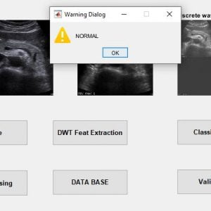
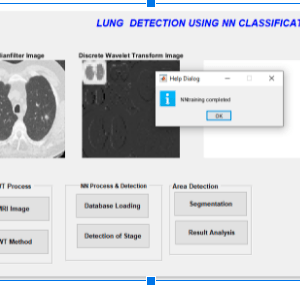
Proposed Method
- DWT and GLCM Features
- NN Classifier
- K-means Clustering
Advantages
- The segmentation algorithm Proves to be simple and effective
- The greyscale Co-occurrence matrix performed well in NN
- Better texture and edge representation
- Segmentation provides better clustering efficiency
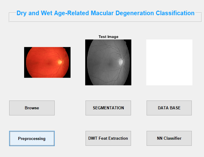
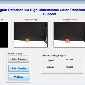
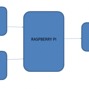
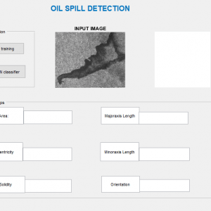
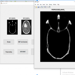
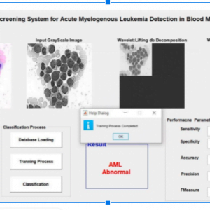
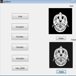
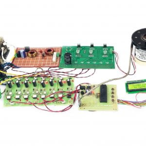
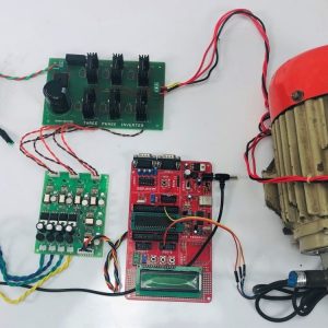
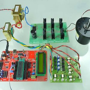
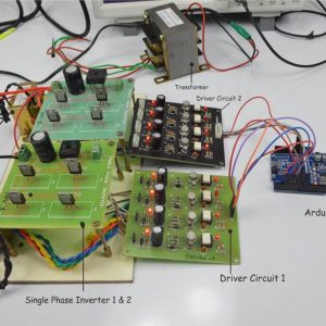
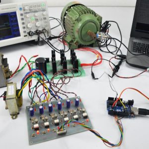
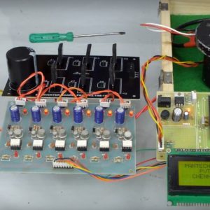
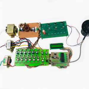
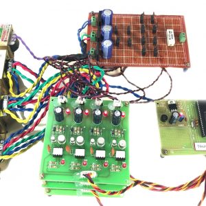
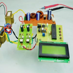
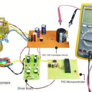
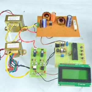
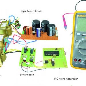
Block Diagram

Hardware Requirements
- system
- 4 GB of RAM
- 500 GB of Hard disk
Software Requirement
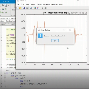
- MATLAB 2014a
Lung Nodule Detection in X ray Images using Image Processing
REFERENCES
- [1] Upadhyay, Y. and Wasson, V. 2014. “Analysis of Liver MR Images for Cancer Detection using Genetic Algorithm”. International Journal of Engineering Research and General Science. Vol.2, No.4, PP: 730-737.
- [2] Kumar, P. Bhalerao, S. 2014. “Detection of Tumor in Liver Using Image Segmentation and Registration Technique”. IOSR Journal of Electronics and Communication Engineering (IOSR-JECE). Vo.9, No.2, PP: 110-115.
- [3] Selle, D.; Spindler, W.; Preim, B. and Peitgen, H. O. 2000. “Mathematical Methods in Medical Imaging: Analysis of Vascular Structures for Liver Surgery Planning”. PP: 1-21.
- [4] Zimmer, C. and Olivo-Marin, J. C. 2005. “Coupled Parametric Active Contours”. Transactions on Pattern Analysis and Machine Intelligence. Vol.27, No.11, PP: 1838-1841.
- [5] Chitra, S. and Balakrishnan, G. 2012. “Comparative Study for Two-Color Spaces HSCbCr and YCbCr in Skin Color Detection”. Applied Mathematical Sciences. Vol.6, No.85, PP: 4229 – 4238.


Customer Reviews
There are no reviews yet.