


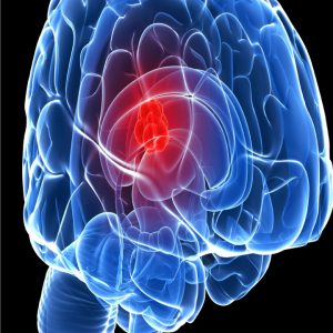
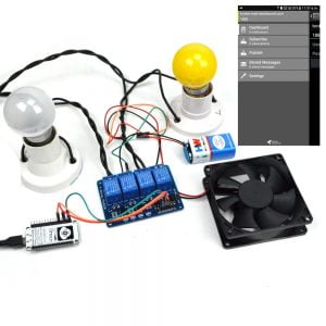
Description
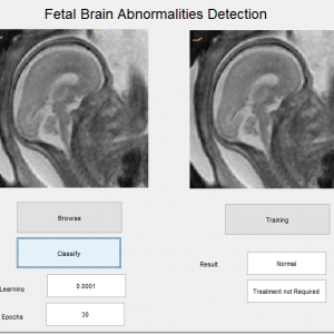
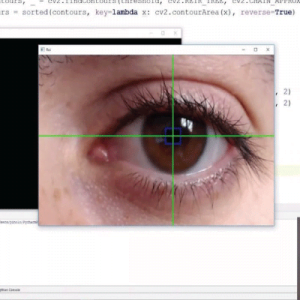
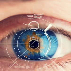
Diabetic Retinopathy Detection using Groud Truth Segmentation
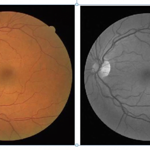
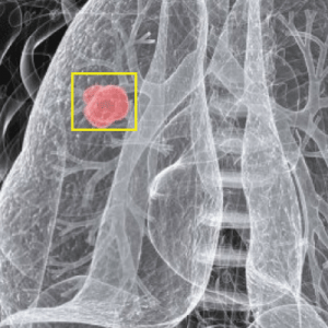
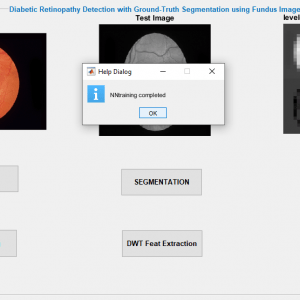
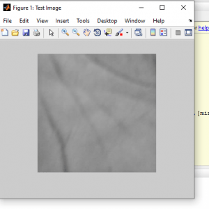
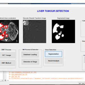
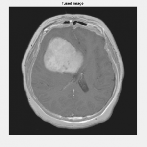
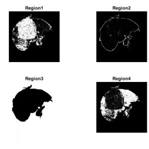
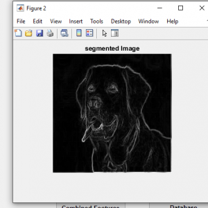
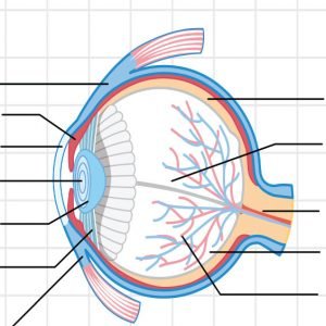
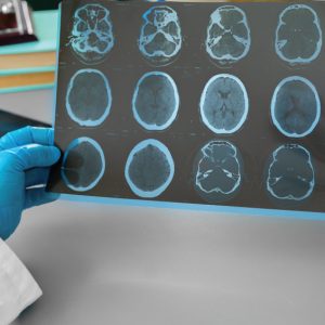
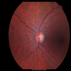
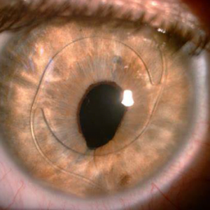
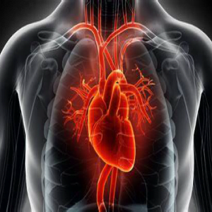
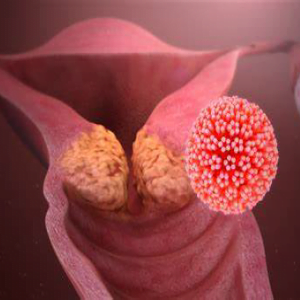
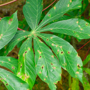
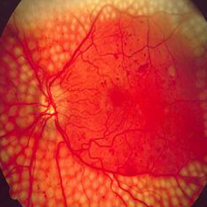
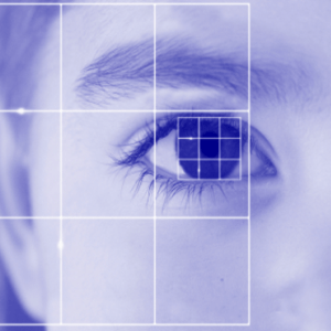
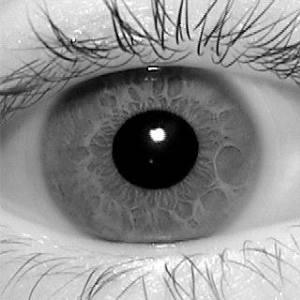
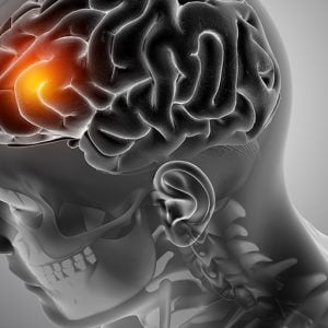
Digital images are obtained from the retina and graded by trained professionals. The progression of diabetic retinopathy is assessed by its severity, which in turn determines the frequency of examinations. However, a significant shortage of professional observers has prompted computer-assisted monitoring. The condition of the vascular network of the human eye is an important diagnostic factor in retinopathy. The Project proposes Retinal image analysis through efficient detection of vessels and exudates for retinal vasculature disorder analysis. It plays important role in the detection of some diseases in the early stages, such as diabetes, which can be performed by comparing the states of retinal blood vessels.
Introduction
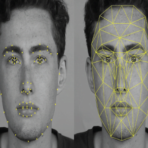
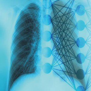
A convolution neural network (CNN) is a type of artificial neural network used in image recognition and processing that is specifically designed to process pixel data.
The layers of a CNN consist of an input layer, an output layer, and a hidden layer that includes multiple convolution layers, pooling layers fully connected layers, and normalization layers.
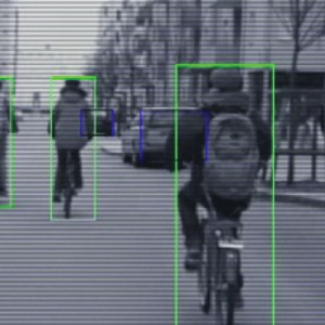
One deep learning approach, regions with convolution neural networks (R-CNN), combines rectangular?region?proposals with convolution neural network features.?R-CNN?is a two-stage detection algorithm. The first stage identifies a subset of regions in an image that might contain an object.
Existing Systems
- Edge detection methods
- Segmentation like simple and global thresholding algorithm
- Retinal Grading Algorithm (RGA)
Drawbacks of Existing System
- Difficulties are there to find the optimal gradient
- In this method, intrinsic characteristics of retinal images make the blood vessel detection process difficult.
- Poor Edge detection.
- Not possible to cluster a Fundus Exudates.
Proposed Method
- The combination of multi-structure morphological process and Segmentation technique is used effectively for retinal vessel detection to identify diabetic retinopathy using a neural network.
Advantages
- The developed method shows a high detection rate and enough robustness against the illumination change.
- Better texture and edge representation
- High accuracy
- Simple process with less time
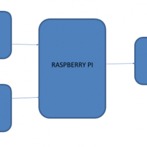
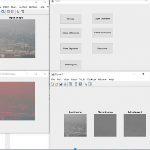
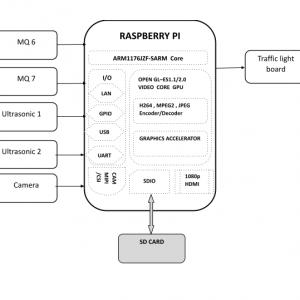
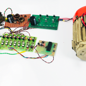
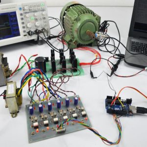
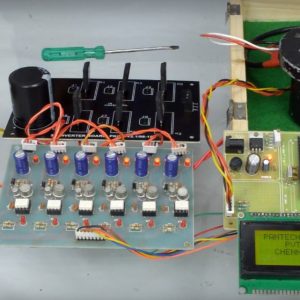
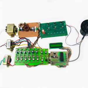
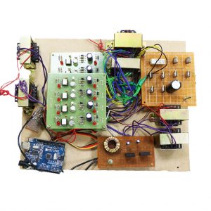
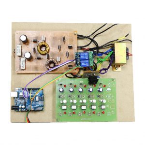
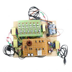
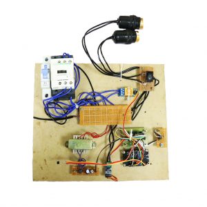
Block Diagram

Diabetic Retinopathy Detection using Groud Truth Segmentation
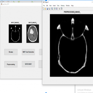
PREPROCESSING:
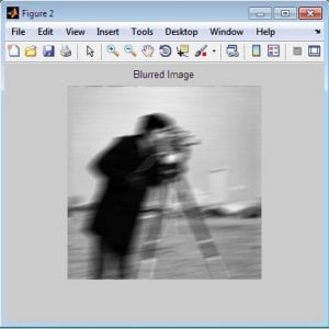
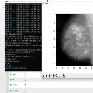
Image restoration is the operation of taking a corrupted/noisy image and estimating the clean original image. Corruption may come in many forms such as motion blur, noise, and camera misfocus.? Image restoration is different from image enhancement in that the latter is designed to emphasize features of the image that make the image more pleasing to the observer, but not necessarily to produce realistic data from a scientific point of view. Image enhancement techniques (like contrast stretching or de-blurring by the nearest neighbor procedure) provided by “Imaging packages” use no a priori model of the process that created the image.? With image, enhancement noise can be effectively be removed by sacrificing some resolution, but this is not acceptable in many applications. In a Fluorescence Microscope resolution in the z-direction is as bad as it is. More advanced image processing techniques must be applied to recover the object.? De-Convolution is an example of an image restoration method. It is capable of Increasing resolution, especially in the axial direction removing noise increasing contrast.
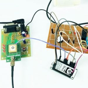
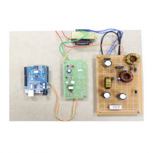
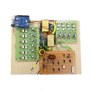
Requirement Specifications
Hardware Requirements
- system
- 4 GB of RAM
- 500 GB of Hard disk
Software Requirement
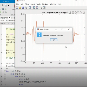
- MATLAB 2014a and above
Diabetic Retinopathy Detection using Groud Truth Segmentation
REFERENCES
[1] M. E. Martinez-Perez, A. D. Hughes, S. A. Thom, and K. H. Parker,?Improvement of a retinal blood vessel segmentation method using the insight segmentation and registration toolkit (ITK),? in Proc. IEEE 29th Annu. Int. Conf. EMBS. Lyon, IA, France, 2007, pp. 892? 895.
[2] S. Dua, N. Kandiraju, and H.W. Thompson, ?Design and implementation of a unique blood-vessel detection algorithm towards early diagnosis of diabetic retinopathy,? in Proc. IEEE Int. Conf. in Inf. Technol., Coding Comput., 2005, pp. 26? 31.
[3] A.M.Mendonc?a and A. Campilho, ?Segmentation of retinal blood vessels by combining the detection of centerlines and morphological reconstruction,?IEEE Trans. Med. Imag., vol. 25, no. 9, pp. 1200?1213, Sep. 2006.
[4] A. Aquino, M. E. Geg?ndez-Arias, and D. Mar?n, ?Detecting the optic disc boundary in digital fundus images using morphological, edge detection, and feature extraction techniques,? IEEE Trans. Med. Imag., vol. 29, no. 11, pp. 1860?1869, Nov. 2010.
[5] M. Lalonde, M. Beaulieu, and L. Gagnon, ?Fast and robust optic disc detection using pyramidal decomposition and Hausdorff-based template matching,? IEEE Trans. Med. Imag., vol. 20, no. 11, pp. 1193?1200, Nov. 2001


Customer Reviews
There are no reviews yet.