


Description
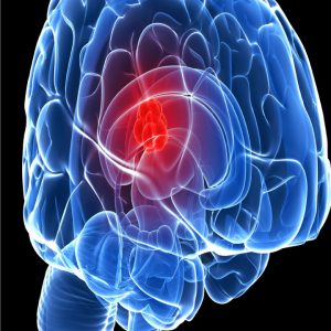
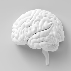
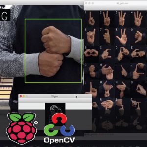
Brain Tumour Segmentation using SFCM & CNN
Abstract
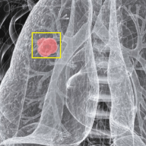
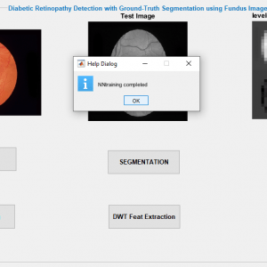
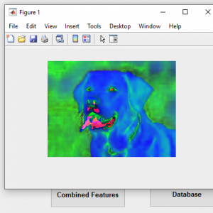
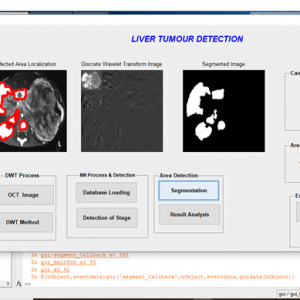
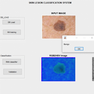
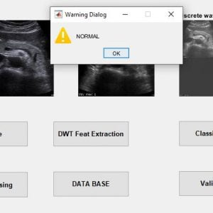
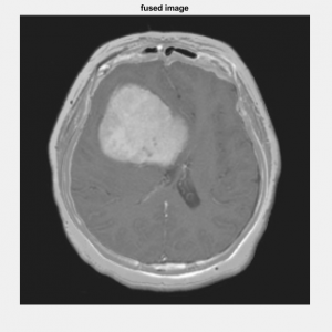
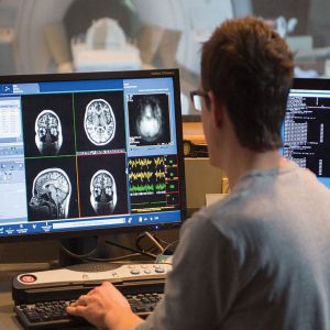
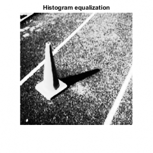
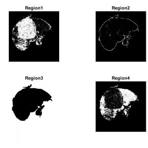
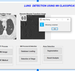
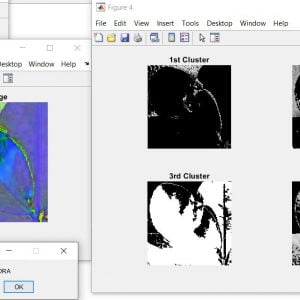
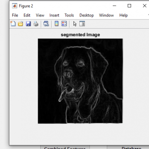
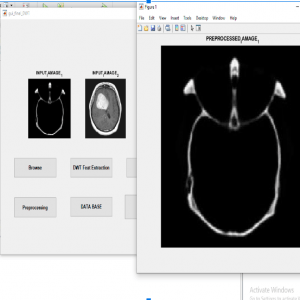
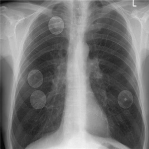
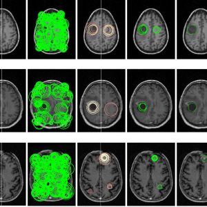
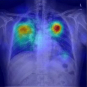
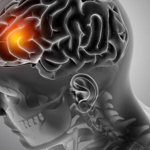
Automatic defects detection in MR images is very important in many diagnostic and therapeutic applications. Because of the high quantity of data in MR images and blurred boundaries, tumor segmentation and classification is very hard. This work has introduced one automatic brain tumor detection method to increase the accuracy and yield and decrease the diagnosis time. The goal is to classify the tissues into three classes normal, begin, and malignant. . In MR images, the amount of data is too much for manual interpretation and analysis. During the past few years, brain tumor segmentation in magnetic resonance imaging (MRI) has become an emergent research area in the field of the medical imaging system. Accurate detection of the size and location of brain tumor plays a vital role in the diagnosis of tumors. The diagnosis method consists of four stages, pre-processing of MR images, feature extraction, and classification. After histogram equalization of the image, the features are extracted based on Dual-Tree Complex wavelet transformation (DTCWT). In the last stage, Convolutional neural Networks (CNN) are employed to classify the Normal and abnormal brain. An efficient algorithm is proposed for tumor detection based on the Spatial Fuzzy C-Means Clustering.
INTRODUCTION:
Brain tumor incidence is a significant contributing factor to the global death rate. According to the Cure Brain Cancer Foundation, brain tumors kill more people under 40 in Australia than any other cancer. In addition, its survival rates are low and have not changed significantly in 30 years, despite the remarkable increase in the survival rate of other types of cancer in Australia.
Existing method
- Partial derivatives
- Wavelet-based denoising
- Thresholding and K mean clustering methods for segmentation
Drawbacks
- Loss of edge details
- In wavelet denoising, failure to detect edge details at curved region.
- K means – It is not suitable for all lighting conditions of images
- Difficult to measure the cluster quality
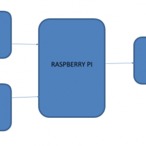
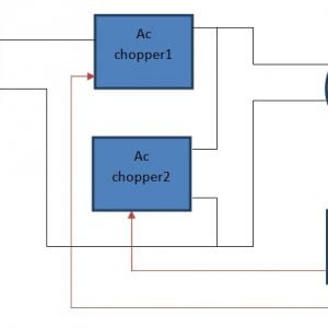
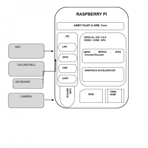
Block Diagram

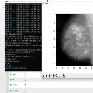
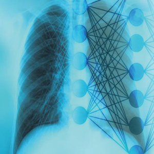
The architecture of a CNN:

All CNN networks have four layers
Brain Tumour Segmentation using SFCM & CNN
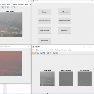
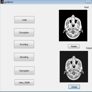
SOFTWARE DESCRIPTION
MATLAB
MATLAB is a high-performance language for technical computing. It integrates computation, visualization, and programming in an easy-to-use environment where problems and solutions are expressed in familiar mathematical notation. Typical uses include:
- Math and computation
- Algorithm development
- Modeling, simulation, and prototyping
- Data analysis, exploration, and visualization
- Scientific and engineering graphics
- Application development, including graphical user interface building
MATLAB is an interactive system whose basic data element is an array that does not require dimensioning. This allows you to solve many technical computing problems, especially those with matrix and vector formulations, in a fraction of the time it would take to write a program in a scalar noninteractive language such as C or FORTRAN.
The name MATLAB stands for matrix laboratory. MATLAB was originally written to provide easy access to matrix software developed by the LINPACK and EISPACK projects. Today, MATLAB uses software developed by the LAPACK and ARPACK projects, which together represent the state-of-the-art in software for matrix computation.
MATLAB has evolved over a period of years with input from many users. In university environments, it is the standard instructional tool for introductory and advanced courses in mathematics, engineering, and science. In industry, MATLAB is the tool of choice for high-productivity research, development, and analysis.
MATLAB features a family of application-specific solutions called toolboxes. Very important to most users of MATLAB, toolboxes allow you to learn and apply specialized technology. Toolboxes are comprehensive collections of MATLAB functions (M-files) that extend the MATLAB environment to solve particular classes of problems. Areas in which toolboxes are available include signal processing, control systems, neural networks, fuzzy logic, wavelets, simulation, and many others.
Conclusion:
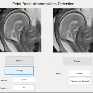
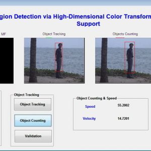
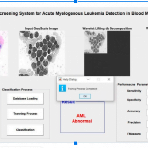
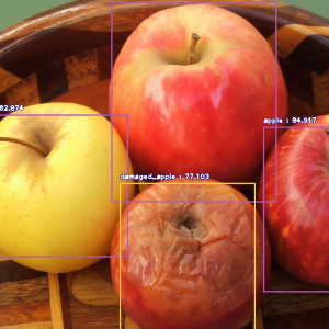
Thus the defects in the MRI images are identified with the help of image processing, in that the machine learning concept called neural network is used to identify the tumor in the image. In the test image, the features are extracted to classify the image. The same features will be extracted in the data set image, by these set of features we can classify the input image as normal or abnormal. Based on the training of the convolutional neural network, the detection is achieved.
Brain Tumour Segmentation using SFCM & CNN
References:
[1] Ardizzone. E, Pirrone. R, and Orazio. O.G, “Fuzzy C-Means Segmentation on Brain MR Slices Corrupted by RF-Inhomogeneity,” In Proc. The 7th international workshop on Fuzzy Logic and Applications: Applications of Fuzzy Sets Theory, WILF ’07, Springer- Verlag, pp: 378-384, 2007.
[2] Binary. P.M, Ashtiyani.M, and Asadi.S, “MRI Segmentation Using Fuzzy C-means Clustering Algorithm Basis Neural Network,” In Proc. ICTT A 3rdInternationai Conference on Information and Communication Technologies: From Theory to Applications, pp: 1-5, 2008.
[3] Sikka. K, Sinha. N, Singh. P.K, and “A fully automated algorithm under modified FCM framework for improved brain MR image segmentation,” Magn. Reson. Imag, vol. 27, pp. 994–1004, Jul. 2009.
[4] Xiao. K, Ho. S.H, and Bargiela. B, “Automatic Brain MRI Segmentation Scheme Based on Feature Weighting Factors Selection on Fuzzy C means Clustering Algorithms with Gaussian Smoothing,” International Journal of Computational Intelligence in Bioinformatics and Systems Biology(3):316-331,2009.


Customer Reviews
There are no reviews yet.