

Description
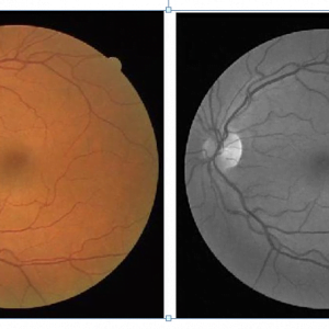
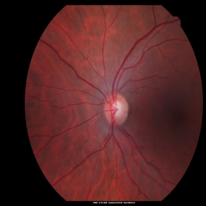
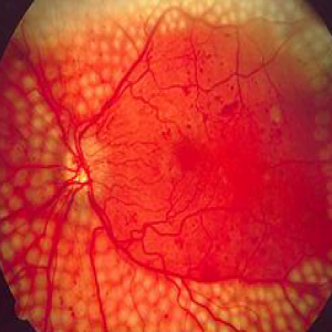
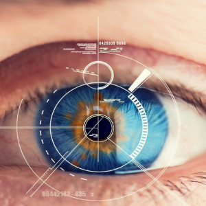
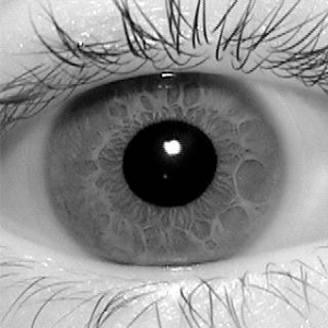
Blood Vessel Segmentation in Retinal Images using Matlab
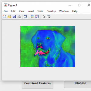
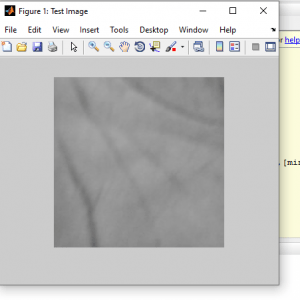
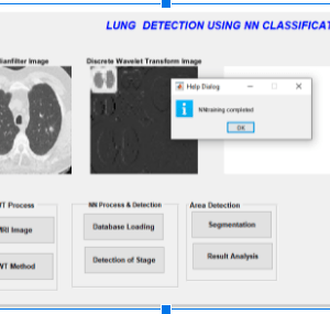
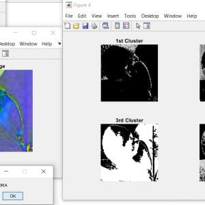
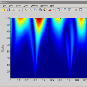
In Our Project, Feature vectors are composed of the pixel?s intensity and continuous two-dimensional stationary wavelet transform responses taken at multiple scales. The Stationary wavelet is capable of turning to specific frequencies, thus allowing noise filtering and vessel enhancement in a single step. Blood Vessel Segmentation in Retinal Images using Matlab
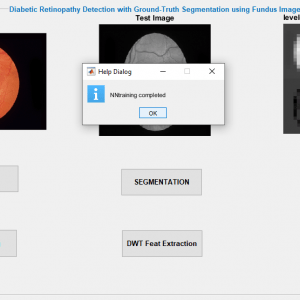
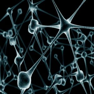
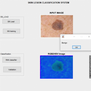
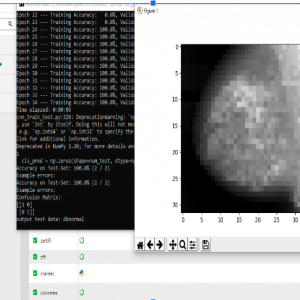
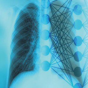
We use a neural network on training with class-conditional probability density functions (likelihoods) described as Gaussian mixtures, yielding a fast classification, while being able to model complex decision surfaces and compare its performance with the linear minimum squared error classifier.
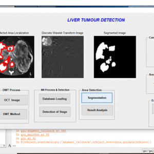
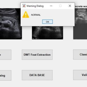
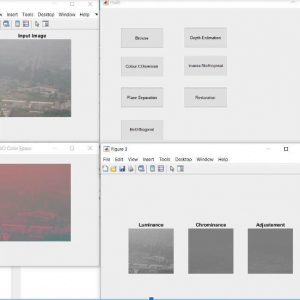
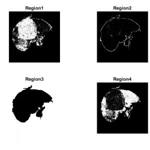
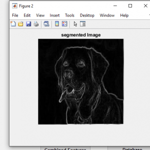
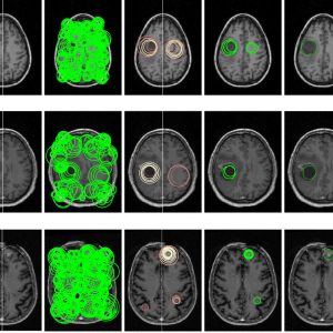
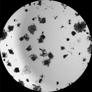
To implement an effective algorithm based on Morphological process and Segmentation Techniques to detect the Retina vessels and Exudates from an eye fundus image. The combination of multi structure morphological process and Segmentation technique is used effectively for retinal vessel and exudates detection here. The modules made here are 1. Retina Blood Vessels Detection in which Plane separation, Contrast Enhancement, Morphological Process are done under this module. 2. Exudates Detection in which Segmentation Technique is used.
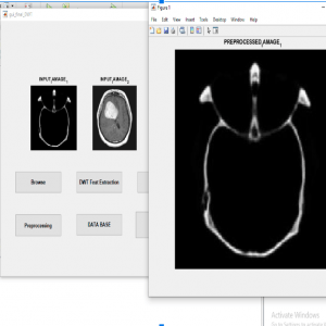
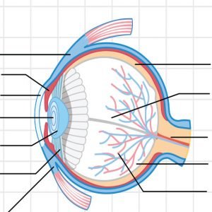
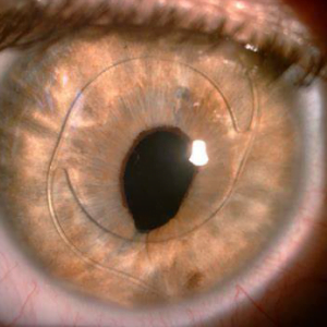
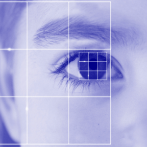
In an automated retinal image analysis system, exact detection of optic disc in colour retinal images is a significant task. Detection of the same is the prerequisite for the segmentation of other normal and pathological features in the retina.. It is seen that optic nerves and blood vessels emerge into the retina through optic disc. Therefore it is also called the blind spot. From patient to patient the size of optic disc varies, but its diameter always lies between 80 and 100 pixels in a standard fundus images. Analysis in medical images is a multi-disciplinary research area, in which image processing, machine learning pattern recognition and computer visualization are covered. Ophthalmologists interprets and analyses the retinal images visually to diagnose various
Pathologies in the retina like Diabetic Retinopathy (DR). In order to make their work more easier retinal image analysis system can be developed to make the diagnosis more efficiently. DR is the most common eye complication in diabetes is Diabetic Retinopathy.
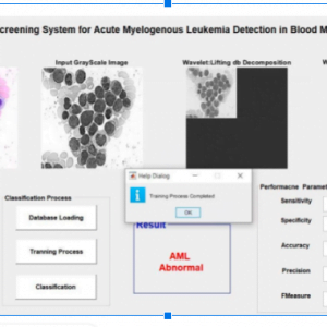
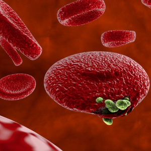
DR is globally the primary cause of visual impairment and causing blindness in diabetic patients. Diabetic patients have to be screened for early detection and timely treatment of diabetic eye diseases which can significantly reduce the risk of vision loss. Reviewing vast number of images by the physicians is time consuming and costly. Several retinal abnormalities including micro aneurysms, haemorrhagess, hard exudates and cotton wool spots are caused due to DR. Hard exudates are yellowish intra retinal deposits, made up of serum lipoproteins. Exudates are formed when lipid or fat leaks from abnormal blood vessels. Vision loss can occur if the exudates extend into the macular area. This paper investigates the application of Morphological approaches for detection of exudates in retinal images and compared with the normal retinal images mainly for the detection of exudates. Blood Vessel Segmentation in Retinal Images using Matlab


Customer Reviews
There are no reviews yet.