


Description
Blood Cell Classification In MicroScopic Images
Abstract:
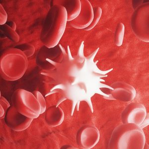
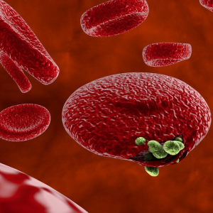
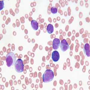
The Blood cells white, red, and platelets are important parts of the immune system. These cells help fight infections by attacking bacteria, viruses, and germs that invade the body. White blood cells originate in the bone marrow but circulate throughout the bloodstream, while red blood cell helps transport oxygen to our body and platelets are tiny blood cells that help your body form clots to stop bleeding. Accurate counting of those may require a laboratory testing procedure that is not usual for everyone. Generating codes that will help count blood cells that produce accurate responses via images gives relief to this problem. In this study, the images were processed and a blob detection algorithm was used to detect and differentiate RBCs from WBCs, and PLATELETs. A cell counting method was also used to provide an actual count of the RBCs, WBCs and PLATELETs detected. The automation comes with a graphical user interface backed up by a working database system to keep the records of the users (e.g. patients, respondents). The performance of the system was statistically described as accurate compared to the manual method of counting. Results show an accuracy of 100% for platelet, 96.32% for RBCs and 98.5% for WBCs. Hence, the proposed system can benchmark with the manual methods of detection and counting of PLATELETs, RBCs, and WBCs in blood samples.
Introduction
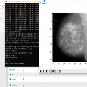
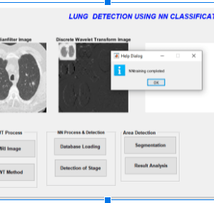
The main objective is to have blood cells containing red blood cells (RBC)and white blood cells and platelet. The RBC carries oxygen from the lungs. The WBCs help fight infection and platelets are parts of CELL ? that the body uses for clotting. All BLOOD CELL are produced in the bone marrow. in this, we use a deep learning technique to classify the cell and count the blood cell. if the person is infected with diseases like dung, malaria cholera, etc it will analyze the blood cells with the help of the neural network technique.
IN this we using preprocessing, dwt, and GLCM? these are techniques that we used and it comes under image processing.? Hemoglobin is an important protein in the red blood cells that carries oxygen from the lungs to all parts of our body.
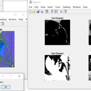
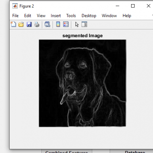
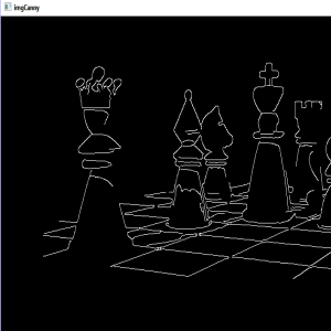
This we are using blob detection to classify the images.
Existing system
- k-means clustering?
- SUPPORT VECTOR MACHINE?
- THRESHOLDING METHODS
Drawbacks:
- It is difficult to do it in machine learning.
- It doesn’t get an accurate rate correctly
Proposed system:
- Pre-processing?
- Dwt
- Glam feature extraction
- Neural network
Advantage:
- It easily detects the blood cell and classifies the counting.
- It easily identifies the blood cells using microscopic images.
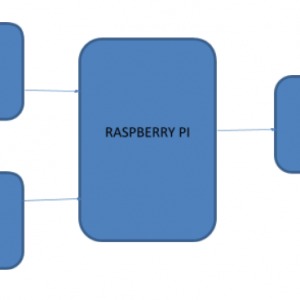
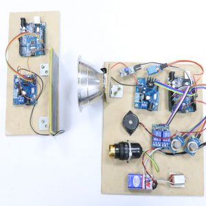
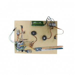
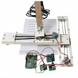
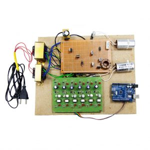
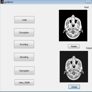
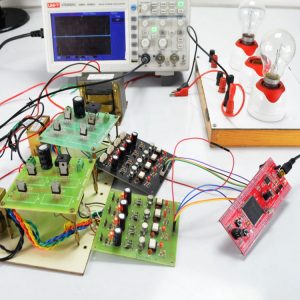
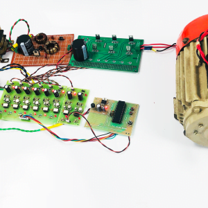
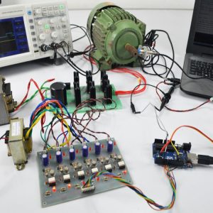
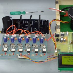
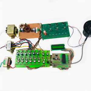
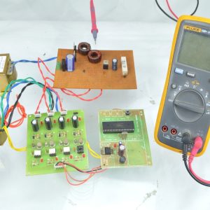
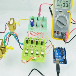
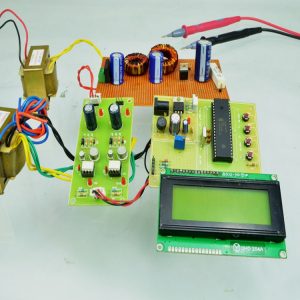
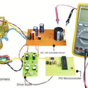
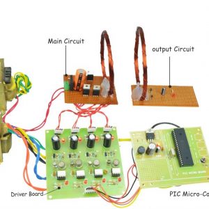
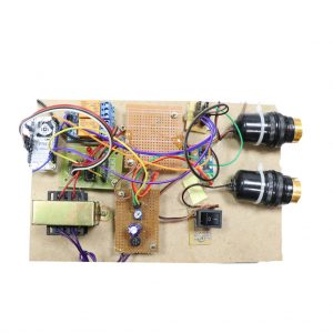
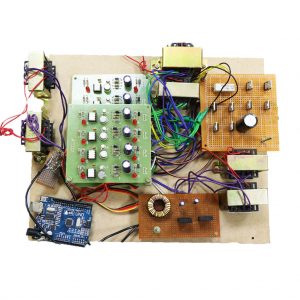
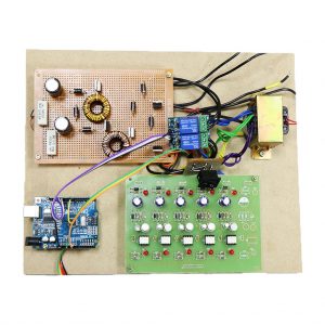
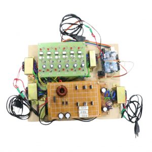
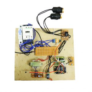
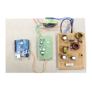
BLOCK DIAGRAM

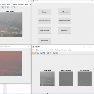
PREPROCESSING
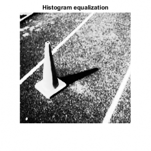
Digital Image Processing. Digital image processing deals with? the manipulation of digital images through a digital computer. It is a subfield of signals and systems but focuses particularly on images.DIP focuses on developing a computer system that is able to perform?processing?on an? image. The input of that system is a digital?image?and the system process that?image?using an efficient algorithm
It allows a much wider range of algorithms to be applied to the input data and can avoid problems such as the build-up of noise and distortion during processing.
- Importing the image via image acquisition tools;
- Analyzing and manipulating the image;
- Output in which result can be altered image
Image Pre-processing? is a common name for operations with?images?at the lowest level of abstraction. Its input and output are intensity? images.? The aim of?pre-processing?is an improvement on the? image? or data that suppresses unwanted distortions or enhances some images? features important for further processing.
Reference:
[1].G .. C.C.Lim, “Overview of Cancer in Malaysia,” Japanese Jomal of Clinical Oncology, Department of Radiotherapy and Oncology, Hospital Kuala Lumpur, 2002.?
[2].Golub TR, Slonim DK, Tamayo P, et al. Molecular classification of cancer: class discovery and class prediction by gene expression monitoring. Science. 1999;286(5439):531-537.
[3].F.Scotti,?Automatic morphological analysis for acute leukemia identification in peripheral blood microscope images,? in Proc. CIMSA, 2005, pp. 96? 101.
[4].Q. Liao and Y. Deng,?An accurate segmentation method for white blood cell images,? in Proc. IEEE Int. Symp. Biomed. Imaging, Atlanta, GA, USA, 2002, pp. 245? 248.
[5].P. Bamford and B. Lovell,?Method for accurate unsupervised cell nucleus segmentation,? in Proc. Eng. Med. Biol. Soc. Conf., 2001, vol. 3, pp. 2704? 2708.
[6].N. Sinha and A. G. Ramakrishnan,?Blood cell segmentation using EM algorithm,? in Proc. 3rd Indian Conf. Comput. Vis., Graph., 2002, pp. 445? 450.
[7].R. D. Labati, V. Piuri, and F. Scotti, ?ALL-IDB: The acute lymphoblastic leukemia image database for image processing,? in Proc. IEEE ICIP, Brussels, Belgium, Sep. 11?14, 2011, pp. 2045?2048.
[8].M.Sezgin and B. Sankur, ?Survey over image thresholding techniques and quantitative performance evaluation,? J. Electron. Imaging, vol. 13, no. 1, pp. 146?165, Jan. 2004.
[9].K.Nallaperumal and K. Krishnaveni, ?Watershed segmentation of cervical images using multiscale morphological gradient and HSI color space,? Int. J. Imaging Sci. Eng., vol. 2, no. 2, pp. 212? 216, Apr. 2008.
[10].R.Adollah, M. Mashor, N. Nasir, H. Rosline, H. Mahsin, and H. Adilah, ?Blood cell image segmentation: A review,? in Proc. IFMBE. Berlin, Germany: Springer-Verlag, 2008, ch. 39, pp. 141? 144.
[11].F.Scotti,?Robust segmentation and measurement techniques of white cells in blood microscope images,? in Proc. IEEE Conf. Instrum. Meas. Technol., 2006, pp. 43? 48.
[12].C. C. Chang and C. J. Lin,?LIBSVM: A library for support vector machines,? ACM Trans. Intell. Syst. Technol., vol. 2, no. 3, p. 27, Apr. 2011.
[13].S.Mohapatra, D. Patra, and S. Sampath,?Image analysis of blood microscopic images for acute leukemia detection,? in Proc. IEC, 2010, pp. 215? 219.
[14].S.Mohapatra, S. Samanta, D. Patra, and S. Sampath, ?Fuzzy-based blood image segmentation for automated leukemia detection,? in Proc. ICDeCom, 2011, pp. 1? 5.
[15]. S. Mohapatra, D. Patra, and S. Sampath,?Automated cell nucleus segmentation and acute leukemia detection in blood microscopic images,? in Proc. ICSMB, 2010, pp. 49?54.
[16]. R. Rangayyan, Biomedical Image Analysis. Series Title: Biomedical Engineering. Boca Raton, FL, USA: CRC Press, Dec. 2004.
[17] R.Walvick, K. Patel, S. Patwardhan, and A. Dhawan. Classification of melanoma using wavelet-transform-based optimal feature set. In SPIE Medical Imaging 2002: Image Processing, volume 5370, pages 944? 951, 2004.


Customer Reviews
There are no reviews yet.